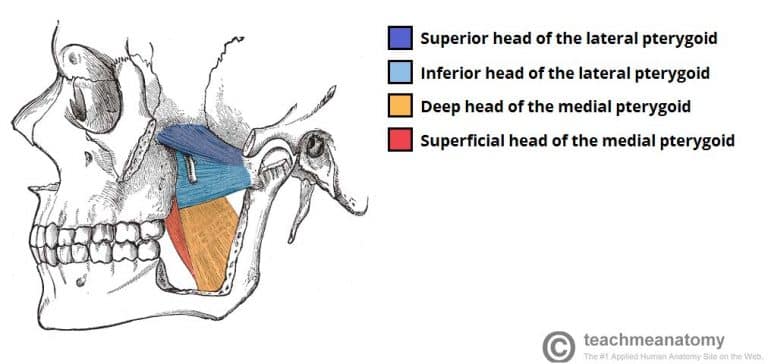
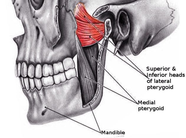
The lateral pterygoid muscle (or external pterygoid muscle) is a muscle of mastication. It has two heads. It lies superior to the medial pterygoid muscle. It is supplied by pterygoid branches of the maxillary artery, and the lateral pterygoid nerve (from the mandibular nerve, CN V3). It depresses and protrudes the mandible. When each muscle works independently, they can move the mandible side to side.
Structure
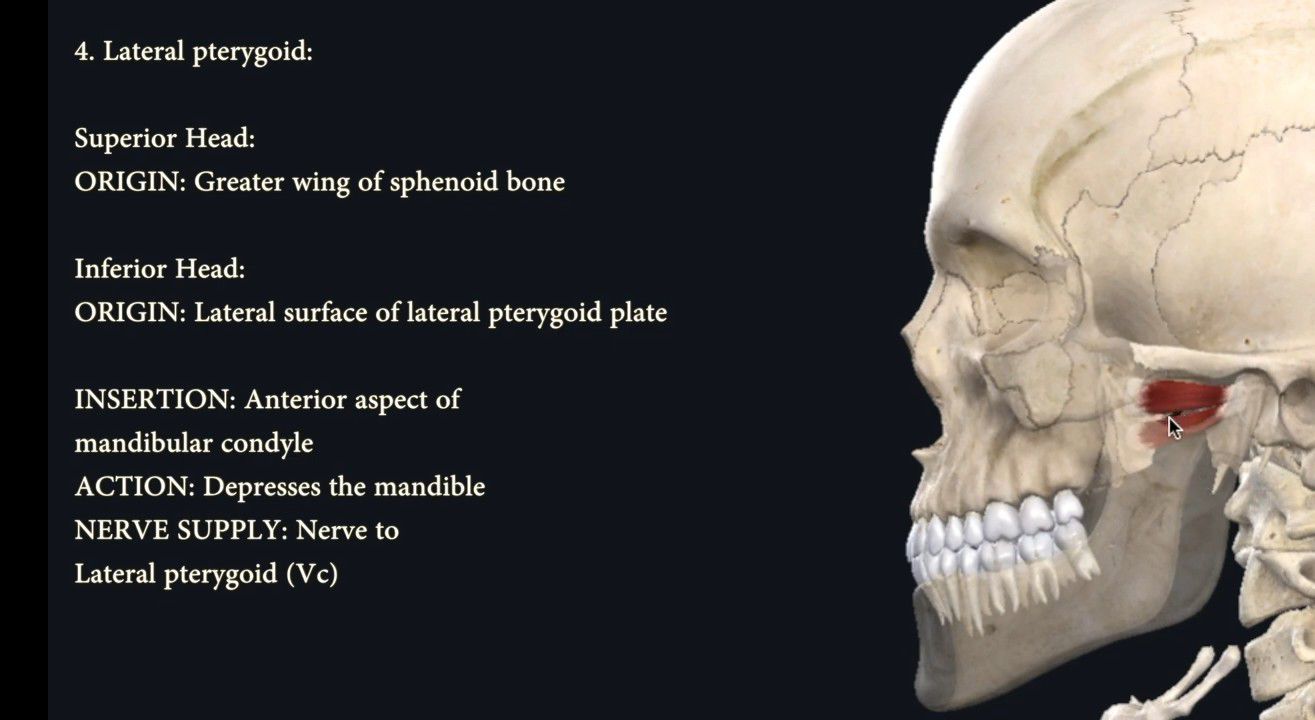
The lateral pterygoid muscle has an upper head and a lower head.
- The upper head originates on the infratemporal surface and infratemporal crest of the greater wing of the sphenoid bone. It inserts onto the articular disc and fibrous capsule of the temporomandibular joint.
- The lower head originates on the lateral surface of the lateral pterygoid plate. It inserts onto the pterygoid fovea at the neck of the condyloid process of the mandible.
It lies superior to the medial pterygoid muscle.
Blood supply
The lateral pterygoid muscle is supplied by pterygoid branches of the maxillary artery.
Nerve supply
The lateral pterygoid muscle is supplied by the lateral pterygoid nerve, a branch of the mandibular nerve (CN V3), itself a branch of the trigeminal nerve (CN V).
Function
The primary function of the lateral pterygoid muscle is to pull the head of the condyle out of the mandibular fossa along the articular eminence to protrude the mandible. A concerted effort of the lateral pterygoid muscles helps in lowering the mandible and opening the jaw. Unilateral action of a lateral pterygoid muscle causes contralateral excursion (a form of mastication), usually performed in concert with the medial pterygoids. When they work independently, they can move the mandible side to side.
Unlike the other three muscles of mastication, the lateral pterygoid alone can assist in depressing the mandible (opening the jaw). At the beginning of this action it is assisted by the digastric, mylohyoid and geniohyoid muscles.
Clinical significance
The lateral pterygoid muscle may be involved in temporomandibular joint dysfunction.
Additional images
References
External links
- lesson4 at The Anatomy Lesson by Wesley Norman (Georgetown University) (musclesofmastication2)
- MedicalMnemonics.com: 70
- "Anatomy diagram: 25420.000-1". Roche Lexicon - illustrated navigator. Elsevier. Archived from the original on 2015-02-26.
- Cross section at tufts.edu